Cell biology is that branch of biology that deals with the study of a plant and an animal cell
Sunday 2 July 2017
Thursday 18 April 2013
knock out technique
A gene knockout (abbreviation: KO) is a genetic technique in which one of an organism's genes is made inoperative ("knocked out" of the organism). Also known as knockout organisms or simply knockouts, they are used in learning about a gene that has been sequenced, but which has an unknown or incompletely known function. Researchers draw inferences from the difference between the knockout organism and normal individuals.
Monday 15 April 2013
Homologous chromosomes
Homologous chromosome
 Homologous chromosomes (also called homologs or homologues) are chromosome pairs of approximately the same length, centromere position, and staining pattern, with genes for the same characteristics at corresponding loci. One homologous chromosome is inherited from the organism's mother; the other from the organism's father.They are usually not identical, but carry the same type of information. Although when Mitosis is occurring the daughter chromosomes are carrying the exact same genetic make up. The product of this is an identical cell- this does however not refer to the occasion where a mutation is occurring.
Homologous Chromosomes
In diploid (2n) organisms, the genome is composed of homologous chromosomes. One chromosome of each homologous pair comes from the mother (called a maternal chromosome) and one comes from the father (paternal chromosome). Homologous chromosomes are involved in the process of meiosis in which they cross over.
Homologous chromosomes are similar but not identical. Each carries the same genes in the same order, but the alleles for each trait may not be the same. In garden peas, for example, the gene for pod colour on the maternal chromosome might be the yellow allele; the gene on the homologous paternal chromosome might be the green allele.
Chromosomes are made of two sister-chromatids, and the chromatids are attatched by centromeres
References:
http://en.wikipedia.org/wiki/Homologous_chromosome
Human Genome Project
The Human Genome Project (HGP)
The Human Genome Project (HGP) is an international scientific research project with a primary goal of determining the sequence of chemical base pairs which make up DNA, and of identifying and mapping the approximately 20,000–25,000 genes of the human genome from both a physical and functional standpoint.[1]
The first official funding for the Project originated with the Department of Energy’s Office of Health and Environmental Research, headed by Charles DeLisi, and was in the Reagan Administration’s 1987 budget submission to the Congress.[2] It subsequently passed both Houses. The Project was planned for 15 years.[3]

In 1990, the two major funding agencies, DOE and NIH, developed a memorandum of understanding in order to coordinate plans, and set the clock for initiation of the Project to 1990.[4] At that time David Galas was Director of the renamed “Office of Biological and Environmental Research” in the U.S. Department of Energy’s Office of Science, and James Watson headed the NIH Genome Program. In 1993 Aristides Patrinos succeeded Galas, and Francis Collins succeeded James Watson, and assumed the role of overall Project Head as Director of the U.S. National Institutes of Health (NIH) National Human Genome Research Institute. A working draft of the genome was announced in 2000 and a complete one in 2003, with further, more detailed analysis still being published.
A parallel project was conducted outside of government by the Celera Corporation, or Celera Genomics, which was formally launched in 1998. Most of the government-sponsored sequencing was performed in universities and research centres from the United States, the United Kingdom, Japan, France, Germany and Spain. Researchers continue to identify protein-coding genes and their functions; the objective is to find disease-causing genes and possibly use the information to develop more specific treatments. It also may be possible to locate patterns in gene expression, which could help physicians glean insight into the body's emergent properties.
While the objective of the Human Genome Project is to understand the genetic makeup of the human species, the project has also focused on several other nonhuman organisms such as Escherichia coli, the fruit fly, and the laboratory mouse. It remains one of the largest single investigative projects in modern science.
The Human Genome Project originally aimed to map the nucleotides contained in a human haploid reference genome (more than three billion). Several groups have announced efforts to extend this to diploid human genomes including the International HapMap Project, Applied Biosystems, Perlegen, Illumina, J. Craig Venter Institute, Personal Genome Project, and Roche-454.
The "genome" of any given individual (except for identical twins and cloned organisms) is unique; mapping "the human genome" involves sequencing multiple variations of each gene.[5] The project did not study the entire DNA found in human cells; some heterochromatic areas (about 8% of the total genome) remain unsequenced.
Among the many social and ethical issues spurred by bio-genetic sciences is a concern regarding bio-genetic warfare (e.g. ethnic bio-weapons targeted towards specific populations).
Benefits
The work on interpretation of genome data is still in its initial stages. It is anticipated that detailed knowledge of the human genome will provide new avenues for advances in medicine and biotechnology. Clear practical results of the project emerged even before the work was finished. For example, a number of companies, such as Myriad Genetics started offering easy ways to administer genetic tests that can show predisposition to a variety of illnesses, including breast cancer, hemostasis disorders, cystic fibrosis, liver diseases and many others. Also, the etiologies for cancers, Alzheimer's disease and other areas of clinical interest are considered likely to benefit from genome information and possibly may lead in the long term to significant advances in their management.
There are also many tangible benefits for biological scientists. For example, a researcher investigating a certain form of cancer may have narrowed down his/her search to a particular gene. By visiting the human genome database on the World Wide Web, this researcher can examine what other scientists have written about this gene, including (potentially) the three-dimensional structure of its product, its function(s), its evolutionary relationships to other human genes, or to genes in mice or yeast or fruit flies, possible detrimental mutations, interactions with other genes, body tissues in which this gene is activated, and diseases associated with this gene or other datatypes.
Further, deeper understanding of the disease processes at the level of molecular biology may determine new therapeutic procedures. Given the established importance of DNA in molecular biology and its central role in determining the fundamental operation of cellular processes, it is likely that expanded knowledge in this area will facilitate medical advances in numerous areas of clinical interest that may not have been possible without them.
The analysis of similarities between DNA sequences from different organisms is also opening new avenues in the study of evolution. In many cases, evolutionary questions can now be framed in terms of molecular biology; indeed, many major evolutionary milestones (the emergence of the ribosome and organelles, the development of embryos with body plans, the vertebrate immune system) can be related to the molecular level. Many questions about the similarities and differences between humans and our closest relatives (the primates, and indeed the other mammals) are expected to be illuminated by the data in this project.
Advantages of Human Genome Project:
Knowledge of the effects of variation of DNA among individuals can revolutionize the ways to diagnose, treat and even prevent a number of diseases that affects the human beings.
It provides clues to the understanding of human biology.
Ethical, legal and social issues
The project's goals included not only identifying all of the approximately 20,000-25,000[31] genes in the human genome, but also to address the ethical, legal, and social issues (ELSI) that might arise from the availability of genetic information. Five percent of the annual budget was allocated to address the ELSI arising from the project.
Debra Harry, Executive Director of the U.S group Indigenous Peoples Council on Biocolonialism (IPCB), says that despite a decade of ELSI funding, the burden of genetics education has fallen on the tribes themselves to understand the motives of Human genome project and its potential impacts on their lives. Meanwhile, the government has been busily funding projects studying indigenous groups without any meaningful consultation with the groups. (See Biopiracy.)[32]
The main criticism of ELSI is the failure to address the conditions raised by population-based research, especially with regard to unique processes for group decision-making and cultural worldviews. Genetic variation research such as HGP is group population research, but most ethical guidelines, according to Harry, focus on individual rights instead of group rights. She says the research represents a clash of culture: indigenous people's life revolves around collectivity and group decision making whereas the Western culture promotes individuality. Harry suggests that one of the challenges of ethical research is to include respect for collective review and decision making, while also upholding the Western model of individual rights.
Monday 17 December 2012
DNA probe
DNA probe
DNA probes are small segments of DNA which help to detect the presence of a gene of a long DNA sequence, in a biological systems. These DNA probes are prepared for commercial purposes and are believed to be the most sophisticated and sensitive means to identify genes or specific DNA sequences. DNA probes provide commercial avenues for diagnosis of infection diseases, identification of food contaminants for isolation of genes and in other microbiological tests.
It is believed that, these DNA probe assays for variety of purposes will be cleaner, simpler, faster and cheaper than the traditional microbiological tests and are also expected to be hundred fold more sensitive.
The production of DNA probes can be done by any of the following methods. Such as
a) using a template DNA with the help of purified biological enzymes
b) DNA probe of specific sequence can also be obtained by using automated DNA synthesizers
c) DNA probe can also be included in viral DNA and may even multiply in bacteria, thus by this way many copies of DNA probe can be obtained.
However, the DNA probe assay consists of the following steps.
Sample to be tested is treated with detergents and enzymes to remove non DNA components. Then DNA is denatured by low PH.
Single stranded DNA binds on filters and is exposed to excess of DNA probes but only one of which will hybridize.
At the same time unbound DNA is detected by a variety of available methods using florescence and dye etc
Friday 7 December 2012
Retinoblastoma
Retinoblastoma - (Reh-tin-oh-blast-oma)
- Retinoblastoma is a rare, cancerous tumor of a part of the eye called the retina
- Retinoblastoma - (Reh-tin-oh-blast-oma) is a cancer of one or both eyes which occurs in young children. There are approximately 350 new diagnosed cases per year in the United States. Retinoblastoma affects one in every 15,000 to 30,000 live babies that are born in the United States. Retinoblastoma affects children of all races and both boys and girls
- The retinoblastoma tumor(s) originate in the retina, the light sensitive layer of the eye which enables the eye to see. When the tumors are present in one eye, it is referred to as unilateral retinoblastoma, and when it occurs in both eyes it is referred to as bilateral retinoblastoma. Most cases (75%) involve only one eye (unilateral); the rest (25%) affect both eyes (bilateral). The majority (90%) of retinoblastoma patients have no family history of the disease; only a small percentage of newly diagnosed patients have other family members with retinoblastoma (10%).

- The eye of an adult measures about one inch from the front to the back of the eye; a child's eye measures about three-quarters of one inch.
- The eye has three layers:
1. Sclera - the outer protective white coating of the eye
2. Choroid - the middle layer which contains blood vessels to nourish the eye
3. Retina - the inner layer which contains the nerves that bring information to the brain for seeing
- The cornea is the clear portion of the front of the eye which bends light rays.
- The conjunctiva is a thin tissue which lines the eyelids and the eyeball up to the edge of the cornea.
- The iris is the colored portion of the eye which is made up of a spongy tissue and is an extension of the choroid.
- The pupil is the opening in the iris (black) which allows light into the eye.
- The lens helps focus light rays onto the retina the way a camera lens focuses light onto film; the lens can change shape, or accommodate, to focus on near or distant objects.
- The eye is filled with fluids which help nourish and maintain the pressure within the eye. The anterior chamber, the front portion of the eye between the iris and the cornea, is filled with aqueous humor, a watery fluid which nourishes the lens and mantains the pressure within the eye.
- The back portion of the eye is filled with vitreous humor, a transparent gel.
- The retina is made up of ten layers and contains over one million cells.
- The optic nerve has nerve fibers which transmit information to the brain for interpretation of objects seen.
- The macula is the area of the retina that is responsible for central vision; its central portion is referred to as the fovea and is responsible for the sharpest vision.
- The macula houses the highest concentration of the cones which are responsible for color and sharp vision.
- The rods, which compose the rest of the retina, are more sensitive to light and are responsible for night vision and peripheral vision.
- Attached to the globe of the eye are six muscles which aid in the movement of the eye. Movement of the eye may be caused by one, a few, or all of the muscles working together.
- Causes, incidence, and risk factors
- Retinoblastoma is caused by a mutation in a gene controlling cell division, causing cells to grow out of control and become cancerous.
- In a little over half of the cases, this mutation develops in a child whose family has never had eye cancer.
- Other times the mutation is present in several family members. If the mutation runs in the family, there is a 50% chance that an affected person's children will also have the mutation. They will therefore have a high risk of developing retinoblastoma themselves.
- The cancer generally affects children under the age of 6. It is most commonly diagnosed in children aged 1 - 2 years.
Symptoms - One or both eyes may be affected.
- The pupil may appear white or have white spots.
- A white glow in the eye is often seen in photographs taken with a flash. Instead of the typical "red eye" from the flash, the pupil may appear white or distorted.
- Other symptoms can include
- Crossed eyes
- Double vision
- Eyes that do not align
- Eye pain and redness
- Poor vision
- Differing iris colors in each eye
- If the cancer has spread, bone pain and other symptoms may occur.
Signs and testsThe health care provider will perform a complete physical exam, including an eye exam. The following tests may be done: - Bone marrow biopsy and cerebrospinal fluid examination in the case of more aggressive tumors
- CT scan or MRI of the head
- Eye exam with dilation of the pupil
- Ultrasound of the eye (head and eye echoencephalogram)
Treatment - Treatment options depend on the size and location of the tumor.
- Small tumors may be treated by laser surgery or cryotherapy.
- Radiation is used for both local tumor and for larger tumors.
- Chemotherapy may be needed if the tumor has spread beyond the eye.
- The eye may need to be removed (a procedure called enucleation) if the tumor does not respond to other treatments. In some cases, it may be the first treatment.
Expectations (prognosis) - If the cancer has not spread beyond the eye, almost all patients can be cured. A cure, however, may require aggressive treatment and even removal of the eye in order to be successful.
- If the cancer has spread beyond the eye, the likelihood of a cure is lower and depends on how the tumor has spread.
Complications
Blindness can occur in the affected eye. The tumor can spread to the eye socket through the optic nerve. It may also spread to the brain, lungs, and bones.
Prevention
Genetic counseling can help families understand the risk of retinoblastoma. It is especially important when more than one family member has had the disease, or if the retinoblastoma occurs in both eyes. - sources : http://www.ncbi.nlm.nih.gov/pubmedhealth/PMH0002025/
- sources : http://retinoblastoma.com/retinoblastoma/frameset1.htm
Tuesday 4 December 2012
Plant Tissue Culture
Plant Tissue Culture
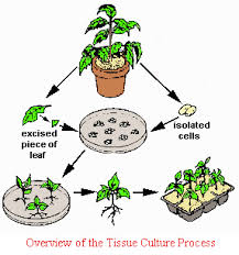
The process by which desirable plants can be grown from any plants part,tissue or cells artificially in the laboratory in an artificially prepared nutrient medium under aseptic condition is known as plant tissue culture.
Historical background of tissue culture:
Haberlandt(1896) was the first person to culture isolated vegetative cells.He was able to maintain the cell in the medium but failed to differentiate it.In 1934 different worker P.R. white,R.s Gautherate and P.Hobegurt were able to grow cambium cells from tobacco stem and carrot root on artificial culture medium.However,this culture failed to differentiate and grew as undifferentiated masses of parenchymatous cells called callus which could be propagated indifinitely by repeated subculturing on fresh culture medium.
Equipments used for Tissue culture:
To get success in vitro culture or micropropagation a laboratory should have following equipments and facilites.They are:
i)pH meter
i)pH meter
ii)Chemical balance
iii)Hot air oven
iv)Centrifuge
v)Auto clave
vi)UV lamp
vii)Shaker
viii)Dissecting microscope
viii)Dissecting microscope
ix)Compound microscope
x)Refrigerator
xi)Laminar air flow cabinet
xii)Essential glass
Methods of Plant Tissue Culture:
a)Nutrient Medium:
Cultured tissue can not synthesize their own food and need an external supply i.e they are heterotroph.The medium or cultured medium used in tissue culture is basal medium.It include following things:
i) Inorganic Nutrients:
It includes all the 16 elements which are essential for normal and healthy life of plants.These elements can be grouped into Macro and micro elements.
Macro elements: C,H,O,P,K,Ca,S,Mg
Micro elements :Zn,Fe,Cu,B,Mn,Mo and Cl
ii)Organic elements :
Micro elements :Zn,Fe,Cu,B,Mn,Mo and Cl
ii)Organic elements :
It includes sucrose,glucose,fructose,carbohydrats and vitamins.
iii)Natural extracts:
natural extract like yeast extract,coconut milk,tomato juice,malt extract are added in the medium.
iv)Growth Hormone:
it includes auxin,cytokinin and gibberellin
Some of the standard media available are Murasbige and Skoog's media,White's media and Nitschs media.
v)Agar:
Agar is a polysaccharide substance obtained from sea weeds which is used to provide solid surface for growth.
b)Sterilization or Aseptic Condition:
The culture must be totally free from microbian contamination.Microbes may enter culture through the ingredients of medium,througn plant organ or explant and through air so that the culture vessel and instrumenti.e glasswares,metal instruments are sterilized by exposure to hot dry air at 160 C to 170 C for 2-4 hours in a hot air oven.
i)Culture Medium:
Culture vessels containing the medium are plugged and autoclaved at 120 C for about 15-20 minutes.
ii)Plant material or Explant:
Plant material or explants are surfacesterilized by using sodium hypochloride or calcium hypochloride solution.After surfacesterilization plant materials are washed 3-4 times in a sterile distill water.
iii)Transfer Arae:
Inoculation is carried out in a laminar air flow cabinet.In this cabinet filterate sterile air flows inside at a constant rate.The flow is unidirectional and makes the cabinet sterile.
To avoid contamination hands and arms are washed with soap and then 95% ethanol.Thus an aseptic environment is maintained for tissue inoculation.
c)Light:
Normally it is not necessary for growth of culture but it plays an important role in inducing differentiation.The intensity and duration of illumination varies from species to species.
d)Temprature:
Generally 25 to 27 degree centigrade is necessary for callus growth.
e)Humidity:
A relative humidity of 70-75% is optimum for the growth of culture.
a)Shoot Culture:
Plant tissue culture in which sterile shoot tips or axillary buds are used as explants is called Shoot culture.
b)Protoplast Culture:
Here,protoplast is used as culture.Somatic hybrid can be produced from protoplast culture.
c)Embryo Culture:
The plant tissue culture in which embroyo is used as explant is known as embryo culture.
d)Anther Culture :
The plant tissue culture in which anther is used as explant is known as Anther culture.By this haploid plant is produced which is of great importance to scientist as mutation can be induced in them.
e) Meristem Culture:
The plant tissue culture in which apical meristem is taken as explant is known as meristem culture.Through this disease resistance plant can be produced.
Rapid asexual or vegetative propagation of plant in vitro is called micro propagation.Large no of plants can be produced throughout the year.
2)Somatic Hybridization:
Fusion of somatic cells in vitro is called somatic hybridisation.Novel hybrid can be produced in sexually incompatible species.
3)Production of Haploid Plants:
Through anther culture haploid plants are produced.It is very important in research point of view as mutation can be induced and detected.
4)Production of Pathogen Free Plants:
Through meristem culture,virus free plants can be produced from diseasesd material.
5)Production of Disease Resistant Varieties:
Many plants are dying due to presence of virus or bacteria.So,plant tissue culture has been able to produce disease resistance variety of plants.
6)Minimize the using space:
Tissue culture can be used to minimize the growing space in commercial nurseries for maintenance of stock plants.
Souce: http://technologysifi.blogspot.in/2010/03/plant-tissue-culture.html
natural extract like yeast extract,coconut milk,tomato juice,malt extract are added in the medium.
iv)Growth Hormone:
it includes auxin,cytokinin and gibberellin
Some of the standard media available are Murasbige and Skoog's media,White's media and Nitschs media.
v)Agar:
Agar is a polysaccharide substance obtained from sea weeds which is used to provide solid surface for growth.
b)Sterilization or Aseptic Condition:
The culture must be totally free from microbian contamination.Microbes may enter culture through the ingredients of medium,througn plant organ or explant and through air so that the culture vessel and instrumenti.e glasswares,metal instruments are sterilized by exposure to hot dry air at 160 C to 170 C for 2-4 hours in a hot air oven.
i)Culture Medium:
Culture vessels containing the medium are plugged and autoclaved at 120 C for about 15-20 minutes.
ii)Plant material or Explant:
Plant material or explants are surfacesterilized by using sodium hypochloride or calcium hypochloride solution.After surfacesterilization plant materials are washed 3-4 times in a sterile distill water.
iii)Transfer Arae:
Inoculation is carried out in a laminar air flow cabinet.In this cabinet filterate sterile air flows inside at a constant rate.The flow is unidirectional and makes the cabinet sterile.
To avoid contamination hands and arms are washed with soap and then 95% ethanol.Thus an aseptic environment is maintained for tissue inoculation.
c)Light:
Normally it is not necessary for growth of culture but it plays an important role in inducing differentiation.The intensity and duration of illumination varies from species to species.
d)Temprature:
Generally 25 to 27 degree centigrade is necessary for callus growth.
e)Humidity:
A relative humidity of 70-75% is optimum for the growth of culture.
Types of plant Tissue Culture:
Plants material used for plant tissue culture is known as explants.On the basis of explants used for plant tissue culture,it is of following type:a)Shoot Culture:
Plant tissue culture in which sterile shoot tips or axillary buds are used as explants is called Shoot culture.
b)Protoplast Culture:
Here,protoplast is used as culture.Somatic hybrid can be produced from protoplast culture.
c)Embryo Culture:
The plant tissue culture in which embroyo is used as explant is known as embryo culture.
d)Anther Culture :
The plant tissue culture in which anther is used as explant is known as Anther culture.By this haploid plant is produced which is of great importance to scientist as mutation can be induced in them.
e) Meristem Culture:
The plant tissue culture in which apical meristem is taken as explant is known as meristem culture.Through this disease resistance plant can be produced.
Applications of Plant Tissue Culture:
1)Micro propagation:Rapid asexual or vegetative propagation of plant in vitro is called micro propagation.Large no of plants can be produced throughout the year.
2)Somatic Hybridization:
Fusion of somatic cells in vitro is called somatic hybridisation.Novel hybrid can be produced in sexually incompatible species.
3)Production of Haploid Plants:
Through anther culture haploid plants are produced.It is very important in research point of view as mutation can be induced and detected.
4)Production of Pathogen Free Plants:
Through meristem culture,virus free plants can be produced from diseasesd material.
5)Production of Disease Resistant Varieties:
Many plants are dying due to presence of virus or bacteria.So,plant tissue culture has been able to produce disease resistance variety of plants.
6)Minimize the using space:
Tissue culture can be used to minimize the growing space in commercial nurseries for maintenance of stock plants.
Souce: http://technologysifi.blogspot.in/2010/03/plant-tissue-culture.html
Subscribe to:
Posts (Atom)